The first ASCRS Main Stage session on Saturday, April 26 was the Get H.I.P. – Cornea session. The Get H.I.P. sessions feature “high impact pearls,” discussed through short, video-based presentations.
Cornea videos were shared in several categories, with discussion following each case. Categories included surgical techniques and tips, surgical or postop complications (and how to manage them), and novel surgical approaches/devices.
Source: ASCRS
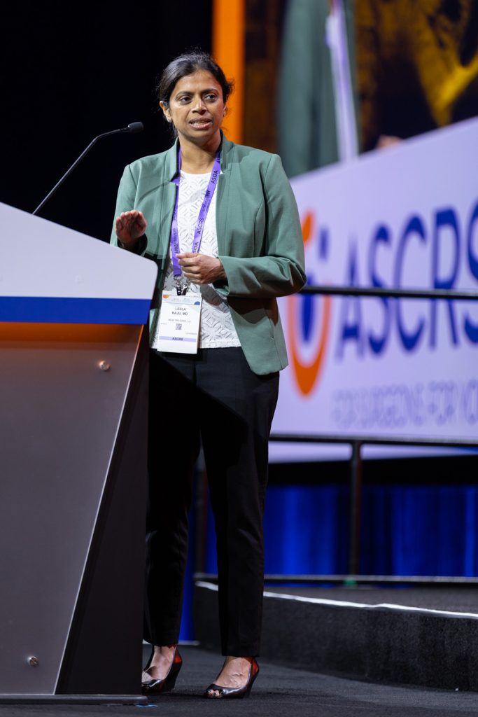
Groove and peel anterior lamellar keratoplasty
Leela Raju, MD, presented the first video of the session, sharing the “groove and peel” technique for anterior lamellar keratoplasty. Her video described that this could work well with keratoconus and patients with anterior stromal scars.
The video showed partial trephination, and a blade was used to deepen the groove in the area where the tissue will be grasped. This can help surgeons be more successful and efficient in anterior lamellar keratoplasty if they’re not high volume, Dr. Raju noted in the video. Forceps were used to pull the anterior lamella.
This leaves a smooth plane and interface, then a 400-micron cap was sutured in place, as with a normal penetrating keratoplasty.
Dr. Raju said at the end of the case that the groove and peel technique (aka “grip it and rip it”) can improve success with anterior lamellar keratoplasty even for a low volume surgeon by using the cornea’s ability to be split, leaving a smooth interface.
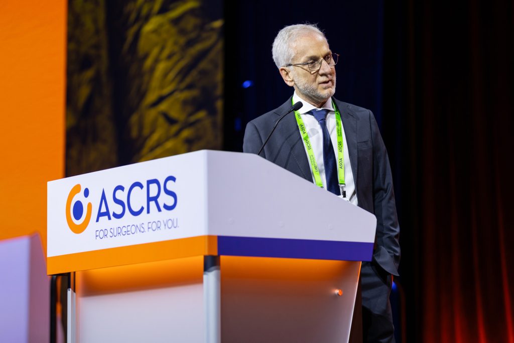
Intrastromal lamellar keratoplasty
Leonardo Gontijo, MD, presented a case using intrastromal lamellar keratoplasty (ILK), which he described as an innovative technique designed for peripheral lamellar reinforcement in cases of extreme corneal thinning. It offers significant advantages over the traditional approach, including shorter surgical time, reduced need for postoperative evaluations, and fewer complications compared to the conventional anterior reinforcement method.
ILK is a straightforward technique, he said. The video noted using a diamond blade incision, then a stromal pocket is created, where a graft is implanted. This eliminates the need for sutures in the lamellar reinforcement process, Dr. Gontijo said.

Source: ASCRS
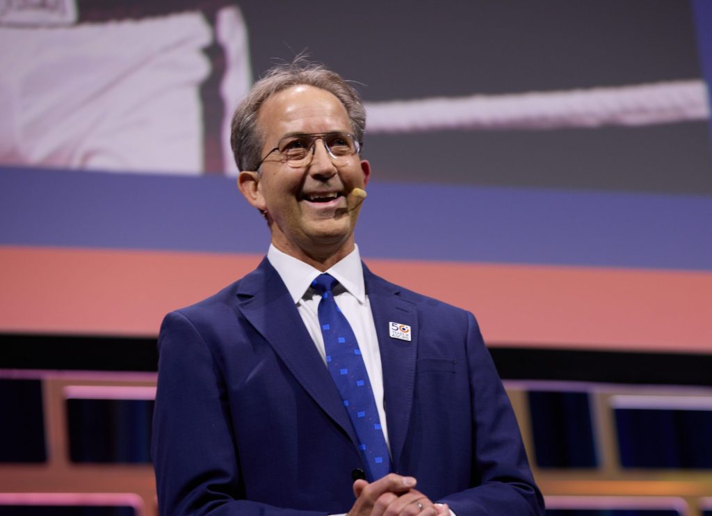
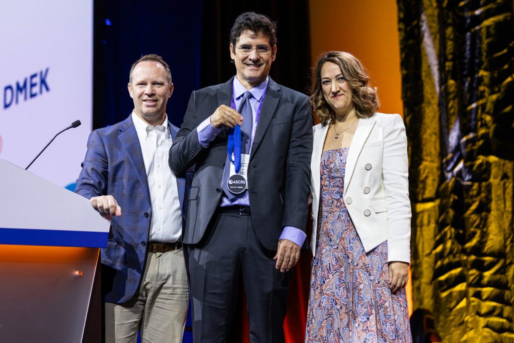
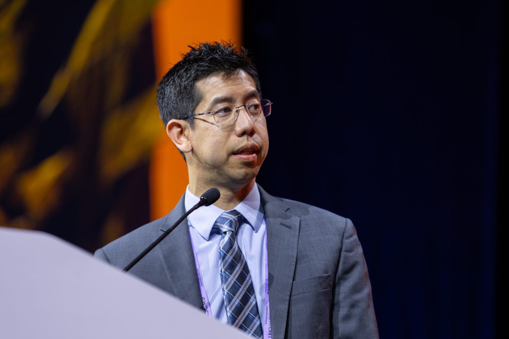
Enhancing descemetorhexis visualization for endothelial keratoplasty
During his video, Albert Cheung, MD, discussed enhancing descemetorhexis visualization for endothelial keratoplasty. He said that adequate removal of Descemet’s membrane optimizes DMEK graft adherence.
He described endothelialectomy with an irrigating cannula and trypan blue staining. Several passes over the desired area are made to ensure adequate treatment. Trypan blue is instilled into the anterior chamber, and after irrigation, the trypan stains the exposed Descemet’s membrane, which enhances visualization. Then you can proceed with descemetorhexis.
Source: ASCRS
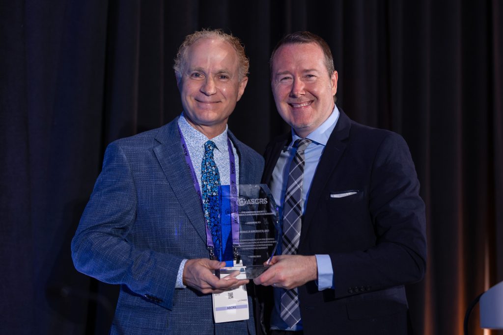
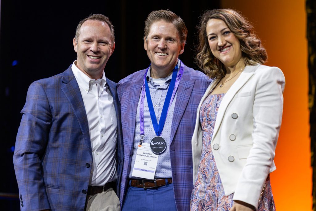
Previous iris melanoma resection: HumanOptics artificial iris
Winston Chamberlain, MD, PhD, shared a case of a previous iris melanoma resection and putting in an artificial iris.
His case was a 65-year-old, who had 40–50% non-useable iris.
He used viscoelastic, then resected the previous iridoplasty. He made a large capsulorhexis in anticipation of the large iris prosthetic in the capsular bag. He used the typical chop technique.
Dr. Chamberlain stained the capsule with trypan blue and placed a CTR in the bag. He measured the dimension of the bag with an intraocular ruler and used a low-profile 3-piece IOL in the bag.
He trephined the iris to the correct size and used a silicone IOL injector to insert the iris. He injected it into the anterior chamber and had the leading edge tucked. You can unravel and tuck portions of the iris under the capsular edges, he said.
Source: ASCRS
Photo Gallery
Relevant disclosures
Chamberlain: None
Cheung: None
Gontijo: None
Raju: None