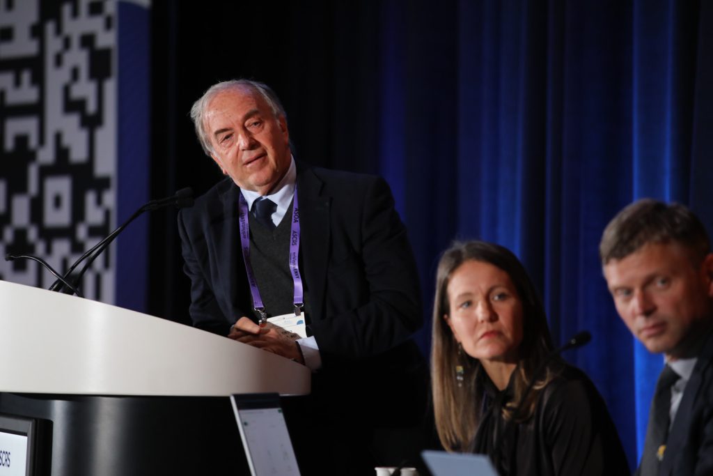
The Winning Pitch Challenge took place at the SightLine at ASCRS meeting on Thursday, April 24. Vance Thompson, MD, and John Berdahl, MD, moderated the session with judges Jim Mazzo, Richard Lindstrom, MD, William Link, PhD, and Ali Bauerlein.
Source: ASCRS
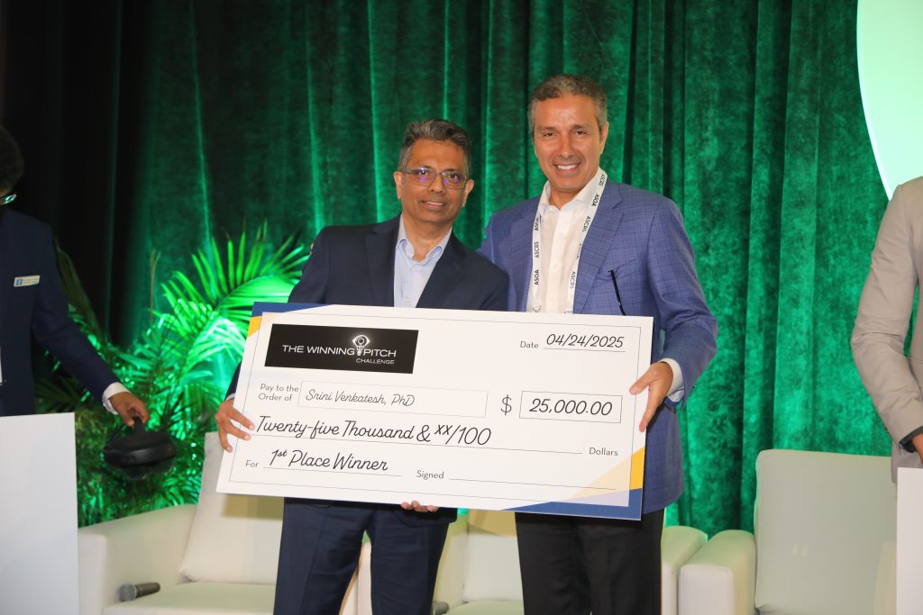
Finalists pitched their ideas to the moderators and judges and were awarded prizes at the end of the session.
First place: Srini Venkatesh, PhD
Second place: Daemon McClunan, MD
Third place: Prasanna Ramesh, MD
Srini Venkatesh, PhD, presented EyeDura, an early-stage seed funded ophthalmic pharmaceutical company developing preservative-free, topical sustained-release eye drops that target both anterior and posterior eye conditions.
There are current limitations for drug delivery in this area, but EyeDura is poised to transform how topical eye drops are used, he said. A single drop of the topical sustained-release (TSR) formulation will deliver therapeutic levels of API for 7 days or longer. The TSR vehicle is compatible with most APIs currently used in ophthalmic products, including small molecules and biologics, Dr. Venkatesh said.
A single eye drop is proven to deliver therapeutic medication levels for multiple days because it is lipid based, has high viscosity, and integrates into the lipid phase of the tear film.
During a blink, the aqueous layer is selectively removed while the lipid layer stays on. The TSR drop resides on the lipid layer, Dr. Venkatesh said, and remains on the surface for extended periods and provides a stable depot.
A probing trial was done with 10 patients with persistent epithelial defects that were refractory to conventional treatment. They were treated with TSR insulin. After 2 drops/week treatment with TSR insulin drops, the epithelial defects healed completely in all 10 patients by day 30. TSR drops were applied only 8 times over 4 weeks. Additionally, he noted looking at the delivery of insulin topical eye drops to the vitreous.
The lead asset is for neurotrophic keratitis, Dr. Venkatesh said.
Daemon McClunan, MD, presented the QMAX by LIQID Medical for the treatment of glaucoma.
Glaucoma is the leading cause of irreversible blindness worldwide. Treatment starts with drops, laser, and/or MIGS, he said. But he also noted the problem of refractory glaucoma and added that 1 in 4 patients will require bleb-forming surgery. Around 1 million of these are done each year. But the problem with a bleb is they are inherently associated with a high rate of complications, high management, and bleb failure.
Dr. McClunan described QMAX as the first and only device to drain excess fluid to a natural fluid reservoir. Advantages are that there is no postop bleb management, costs, and complications. There is unmatched and sustained eye pressure control, and it’s ideal for high and normal tension glaucoma. Dr. McClunan said there is no toxic MMC required, and it’s suitable for inferior placement.
He noted results with trialing this option for severe/refractory glaucoma showing greater than 60% reduction in IOP and greater than 70% reduction in medications.
The surgical technique, Dr. McClunan said, is a 25- to 45-minute procedure that uses an inferior or superior approach. The custom retractor provides clear exposure, and it has a minimally invasive delivery system. He also said there is a rapid postop recovery.
Prasanna Ramesh, MD, presented the Eye MG by MCMI.
This aims to enhance diagnostics, education, and patient engagement using unique 4D live scan imaging technology.
This addresses a couple of problems, he said. The first is that the existing diagnostic tools are difficult for young ophthalmologists to interpret. The eye, being a three-dimensional organ, can cause potential misunderstandings or misinterpretations of its structure when depicted on a two-dimensional surface. The second problem is that fundus scans and OCT are limited in image representations and can be difficult to interpret. For the same patient, interpreting both the fundus scan and the OCT scan requires experience to recognize that they provide the same diagnosis.
The solution is the Eye MG, which Dr. Ramesh said brings 4D life to the 2D scan images. Eye MG is a translational software that enhances the 2D scan images and projects them onto a 4D environment. It is not an imaging tool, he noted.
Eye MG is available with a number of devices, including corneal topography, specular microscopy, ultrasound biomicroscopy, AS-OCT, Scheimpflug imaging, fundus capture device, OCT, and biometry. Eye MG has been designed to be compatible with a wide range of devices and platforms.
The market for this would be ophthalmologists, optometrists, eye institutes, and diagnostic manufacturers and electronic medical records companies, he said.